Causes of eye asymmetry
Facial asymmetry can develop immediately after birth. This means that the condition began while still inside the womb. Another type of disease is pathological, and occurs several months or years after birth.
Anomalies of the skull structure
Incorrect skull structure is inherited. Defects may include various irregularities and depressions:
- the right side is larger than the left, or vice versa;
- the affected area is excessively large;
- sloping forehead or back of the head;
- one cheekbone is larger than the other.
All these factors change facial features, making them asymmetrical.
Developmental disorder of the lower jaw
The most harmless condition is malocclusion. A person develops a jaw that is pushed forward, a disruption in the normal development of wisdom teeth, and a receding or overly large chin. The lower jaw is one of the largest parts of the face, so its violations affect a person’s appearance.
Muscle and connective tissue problems
If a person has weakness in the facial muscles, drooping of the eyelids, cheeks, and eyebrows occurs. Reduced quality of connective tissue contributes to sagging skin. If this condition is common on one side of the face, it will be asymmetrically lower than the other.
A typical picture of muscle and connective tissue disorders is one eye wide open, the other narrow.
If the muscle tissue disorder can be not only on the face, but also on the neck. If it is very tense, the person's head will lean in this direction.
Violation of the structure of the temporomandibular region
If a person has a disorder in the joint between the lower jaw and the temporal bone, movement is blocked when opening the mouth. It is formed unevenly; a person can open first to the right side, then to the left, and vice versa. If the condition is severe enough, the person must first move it, then open it.
Strabismus
Strabismus develops in one or both eyes. Defects form immediately after birth or after some time. The causes are various neurological and ophthalmological primary diseases. The coordinated work of the eyes is disrupted. If one organ of vision decreases in acuity, it gradually turns off its function.
Vision defects
There are various diseases that reduce vision function. Many of them form external changes. A person develops a cataract, a cataract, or a scar on one eye. At the same time, the second eye remains healthy, this forms asymmetrical facial features.
Mechanical damage and inflammatory conditions of nervous tissue
Such diseases lead to swelling. If the nerve is inflamed on only one side, it increases sharply in size. Additionally, a pain syndrome is formed. This is observed with mechanical damage, the motor activity of one side is impaired. A person retains facial expressions only on the healthy side.
Absence of a large number of teeth
If the changes are only on one side, the cheek is pressed inward. The healthy part of the face remains normal. A person's face changes when talking. A lisp and a lack of pronunciation of certain sounds appear.
Bad habits
These include:
- squinting of one eye;
- Constantly chewing gum or foreign objects;
- sleep on one side.
Defects develop gradually. A bad habit affects muscle, connective and bone tissue. The location of muscles and bones relative to each other changes. If a large area is affected, the defect can only be corrected through surgery.
Stroke: causes
One of the most dangerous causes of facial asymmetry and impaired facial expressions is a circulatory disorder in the brain, which often results in a stroke. Depending on the cause that caused it, it can be ischemic (when a cerebral vessel is blocked by a thrombus or embolus), as well as hemorrhagic (when a thinned wall of a cerebral vessel ruptures).
Underlying conditions that increase the risk of stroke:
- cerebral atherosclerosis is one of the main causes of ischemic stroke;
- arterial hypertension - increases the risk of hemorrhagic stroke;
- cerebral aneurysm - sac-like protrusion and thinning of the wall of a cerebral vessel, as a common consequence of prolonged arterial hypertension;
- chronic cerebrovascular accidents;
- obesity and low physical activity.
How to recognize pathology?
The main goal of diagnosing ptosis of the upper eyelid is to establish the cause that led to the development of this disease.
- Assess the position and mobility of the eyelid.
- Assess the symmetry of eye movements.
- Assess eyebrow mobility.
- Determine the size of the eyelid fold.
- Determine the strength of the muscle that lifts the upper eyelid.
- Determine the presence of strabismus, amblyopia.
- Check your vision.
- Measure intraocular pressure.
If ptosis is caused by mechanical damage, the doctor should check the bone structures for damage. To do this, you need to conduct a survey x-ray. If there is a suspicion that ptosis has appeared due to problems with the nervous system, then a computer or magnetic resonance imaging of the brain is performed, and a referral to a neurologist and neurosurgeon is given.
- General clinical blood test. It will show leukocytosis and accelerated erythrocyte sedimentation characteristic of inflammation.
- Biochemistry of blood. This laboratory test will help identify cytolytic enzymes. Their level increases if there is destruction of cells and tissues in the body.
- General ophthalmological examination. As part of it, visual acuity is measured using Sivtsev tables.
- Perimetry. This is a method for determining peripheral vision and the degree of its impairment.
- Ophthalmoscopy. Using a slit lamp, the technique examines the condition of the fundus of the eye, the exit point of the optic nerve, the vascular network, and the anterior and posterior chambers of the eye.
- Magnetic resonance and computed tomography of the head. These instrumental methods help to identify hemorrhages, foci of destruction or neoplasms in the cranial cavity.
- Measurement of cerebrospinal fluid pressure. At the same time, cerebrospinal fluid should be collected for microbiological examination.
We invite you to familiarize yourself with the instructions for using Diklo-F eye drops
Causes
Neuritis of the facial nerve can be caused by various reasons:
- hypothermia. Patients themselves most often say that they are “blown away.” And indeed, neuritis can appear due to the fact that the nerve is caught in the cold or a cold wind blows on the face;
- mechanical compression. Often the nerve is compressed by surrounding tissue, for example, due to a tumor. It happens that the cause of neuritis of the facial nerve is tooth extraction;
- damage. Since the nerve branches are located deep, it is impossible to damage them under normal conditions. But sometimes this happens during an accident, some kind of injury, or during surgery.
- infection, exposure to toxins. It can occur as a secondary disease after an infection enters the body.
Symptoms
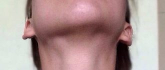
With asymmetry, the right and left sides are different. If they are not natural, the change is observed by 1-2 mm, no more. If the condition is caused by a pathological factor, the asymmetry is visible to the naked eye. Many patients are not satisfied with this, so they turn to surgeons.
The following asymmetrical features are distinguished:
- violation of the location of folds on the face (the area between the lips and nose, folds on the eyelids);
- when smiling, lips curl downwards rather than upwards;
- one-sided smile;
- greater width of one eye compared to the other;
- impaired muscle mobility on one side of the face;
- the formation of discomfort and pain in the damaged area;
- speech defects, lack of pronunciation of certain sounds;
- if a prolapse of connective muscle tissue occurs, the person looks exhausted and tired;
- abnormal location of the palpebral fissure.
Facial asymmetry can not only appear externally, but also lead to pain. In this case, urgent treatment is required. Timely consultation with a doctor reduces the risk of complications.
How to align different halves of the face
If you have identified the fact that the face has significant differences on both sides due to some kind of disease, then to determine the cause, you need to visit a doctor. The specialist will prescribe tests for you, perform an examination and advise the correct treatment.
Asymmetry can occur as a result of drug use. In this case, the face often becomes gray.
To determine the cause of pathological facial asymmetry, you need to visit a doctor. In fact, you may need a number of specialists, but it is best to visit your doctor first.
In this case, you will not waste a lot of time visiting unnecessary medical professionals. The specialist will make a list of doctors you need to visit.
What doctors may be needed for asymmetry:
- Neuropathologist;
- Ophthalmologist;
- Surgeon;
- Orthodontist;
- Dentist.
Once the cause of your illness is discovered, you can undergo treatment and return everything to normal. However, in some cases, additional plastic surgery may be required after treatment.
How to recognize pathology?
It is not difficult to notice the asymmetry of the folds of the face. But recognizing the reason why one eye is smaller or larger than the other is not so easy. In newborns, asymmetrical palpebral fissures are congenital anomalies of the development of facial muscles and skin. But the cause can also be intrauterine and birth injuries to the head, in which hemorrhage occurs in the intracerebral cavities.
Therefore, to diagnose pathology, it is important to know the general history and protocol for pregnancy management. As for adults, the difference in eye size is visible to the naked eye. It is accompanied by concomitant clinical symptoms. It includes headaches, loss of consciousness, neurological disorders, unsteadiness when walking, and the inability to read text or look at pictures.
Blepharospasm: treatment
If your face is distorted due to blepharospasm, you should turn to the following therapeutic methods:
- treatment of the underlying disease that caused spasm of the orbicularis oculi muscle;
- physiotherapy;
- nootropics - drugs that increase oxygen delivery to the brain and improve its functioning;
- If symptoms are severe and other treatment methods are ineffective, botulinum therapy may be prescribed, which effectively relieves muscle spasm.
Diagnostics
Diagnosis of the patient’s condition consists of several stages:
- Anamnesis collection. This is data obtained from the patient or his close relatives. Based on them, the doctor suggests a diagnosis.
- General inspection. Assessment of the condition of skin surfaces and muscle tissue. Doctors identify neurological symptoms, the degree of mobility of facial tissue, the presence of injuries and diseases.
- Lab tests. With the help of testing, an infectious focus and inflammation are identified. A general blood and urine test and blood biochemistry are prescribed.
- Assessing facial symmetry using measuring instruments. The doctor reveals the exact proportions, damage and deformations are detected. If a person has pathological asymmetry, it is formed when the location of parts of the face is disturbed by 3 mm or more.
Based on diagnostic data, the doctor makes a reliable diagnosis. Next, treatment is carried out.
Facial nerve paralysis: causes
Being one of the conditions that can cause the face to become skewed, it requires proper treatment to restore the previous shape of the face. Paralysis is the inability to move any part of the body, in this case, the facial muscles. A number of pathological conditions can lead to this:
- infectious diseases (chicken pox, herpes, rubella) that lead to inflammation of the facial nerve - neuritis;
- inflammation of the middle ear - otitis media;
- traumatic brain injuries;
- inflammation of the meninges - meningitis;
- brain tumors.
Fortunately, all of the serious causes listed above (brain injuries, tumors, meningitis) are rare. But in most cases, the cause of the paralysis cannot be determined, and then doctors make a diagnosis of “idiopathic Bell's palsy.” This condition is the most common cause when a child's face is distorted.
Why does facial asymmetry occur?
There are two main reasons for the development of such a visual deviation: congenital or acquired. These include: • defect in the formation of the temporomandibular joint; • abnormal structure of the skull; • pathology of the formation of the neck muscles (on one side); • general underdevelopment of the lower jaw; • various defects in connective tissue or muscles.
Acquired asymmetry can occur as a result of: • inflammation, injury or pinching of the endings in the facial nerve; • visual defects due to strabismus; • bite pathologies, problems with teeth or jaw; • in the absence of teeth on one half of the jaw; • various facial and jaw injuries, facial bone fractures;
In children, this defect develops with muscular or neurogenic torticollis, congenital shortening on one side of the sternocleidomastoid muscle.
Facial asymmetry also occurs with aging of the entire human body or with age-related diseases.
Symptoms
In the presence of natural asymmetry, the difference in size between the right and left sides of the face is not pronounced (about 2-3 mm), only the overall proportionality is slightly disturbed.
Most often, it is the right half that is larger and wider, and the left half is delicate and has a smoother appearance, but these deviations are not pronounced, so there is no reason to worry.
If there is neuropathy of the facial nerve, then the indicated differences in the symmetry of the face are more visible, and their clinical picture is more pronounced: • in the affected half of the face there is weakness in the facial muscles and it becomes like a mask; • nasolabial and frontal folds are less noticeable; • the palpebral fissure increases;
• the corners of the mouth fall down; • the affected part acquires a pained or crying expression; • it is difficult to move the facial muscles, close your eyes, wrinkle your forehead; • speech and articulation are impaired, it is difficult to eat, because it just falls out of your mouth; • painful sensations are noted in the affected nerve.
Facial asymmetry in children occurs due to muscular torticollis or when lying in a crib on one side for a long time. Most often, part of the face is smoothed, the jaw angle is smaller, the head is tilted towards the affected side, and parts of the face (in the area of the cheek and head) are flatter.
The diagnosis is based on identifying facial asymmetry using a visual examination (presence of pathology of muscles, teeth, nerves), asking the patient about heredity and possible injuries.
Additionally, facial proportions are measured with special instruments, which are expressed in millimeters and degrees. A deviation of more than 3 mm or 5 degrees is considered pathology.
In order to prove the presence of asymmetry in the face, an image of a person’s face is taken, composed of its two right or left halves. The result is two absolutely symmetrical portraits, which often differ from the person’s real face.
We will describe the methods that are used to correct the main parts of the face.
1. Eyebrow asymmetry. The defect develops when the facial nerve, its frontal branch, is damaged. Also, its manifestation is facilitated by hyperfunction of the levator brow muscle. In order for symmetry to be restored, drugs are injected into the muscles (frontal, corrugator brow): Lantox, Botox, Dysport.2. There are numerous muscles around the mouth that work in different directions. Asymmetry around the mouth is corrected by introducing Botox, Lantox into the area of the muscle that depresses the lower lip and the area of the mouth
3. For asymmetry of the eye slits in the presence of the “big eye versus small eye” symptom, medications Lantox, Botox, and Dysport are used. A small dose of botulinum toxin can also be used; the drug is injected into the lower part of the orbicularis oculi muscle, 1 mm away from the edge of the eyelashes.
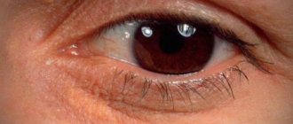
We all understand that the body of any person cannot be absolutely symmetrical, so there is no need to worry about this. But when eye asymmetry occurs, this already indicates the presence of pathology and you should definitely consult a specialist for advice.
When eye asymmetry occurs, the reasons may be the following: 1. The presence of infectious eye diseases, in this case with slight swelling of the eyes becoming larger (for example, barley or adenoviral conjunctivitis). The tumor occurs due to an attack by pathogenic bacteria, which leads to inflammation of the mucous membrane of the eye.
We invite you to familiarize yourself with Prenacid - instructions for use, description, reviews, analogues
Therapy and diagnosis are carried out only by an ophthalmologist, because self-medication can lead to worsening of the condition. 2. Injuries. It should be borne in mind that even a small injury or bruise in the eye area can cause swelling. Treatment of injuries is carried out depending on their formation, but only by a specialist. 3.
If swelling of the eye and a change in its size occurs without a reason, this situation is considered the most dangerous. This defect can lead to neurological disease or even more serious illness. 4. Bulbar syndrome is a dangerous pathology that is associated with the condition of the brain.
At the initial stage of development of the disease, asymmetry of the eyes is noted. It is at this moment that you should consult a doctor to prevent the development of a disease when one of the eyes does not function properly (for example, paralysis). Most often, in parallel with a change in the size of the eye, deformation of the eyelid, incomplete closure of the eyes, and a change in the shape of the eye occur.
Let's draw conclusions: if you have a visually visible difference in the size of your eyes, you should pay attention to the accompanying symptoms (swelling of the eyelid, discharge with purulent contents, redness of the eye mucosa). If the deformity is accompanied by attacks of pain, most often a diagnosis of “neuralgia” can be made.
All pathologies must be diagnosed by a specialist, so do not delay a visit to an ophthalmologist.
Why is one cheek larger than the other (video)
Facial asymmetry can be natural or pathological. If you suspect the second type of this problem, then you need to see a doctor and identify the real cause. If you are sure that asymmetry is harmless, but want to correct it, use our exercises. Take care of your beauty and be healthy!
Comments
+7 Katya 02/10/2018 21:05 I would recommend seeing a doctor, I had this happen when the flux was very swollen.
One cheek was much larger, it couldn’t even go outside. Everything went away after the inflammation subsided, about a week passed. Quote
Update list of comments RSS feed of comments for this entry
Treatment
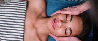
There are various treatment methods. They are selected by the doctor individually for each patient. He, in turn, chooses the most appropriate method:
- Facial correction using cosmetics. If it is necessary to narrow the area, it is darkened. If you expand it, it brightens it up. Corrective foundation creams and pencils are used for this.
- Maxillofacial or plastic surgery. They contact a plastic surgeon, who individually selects the type of operation for the patient.
- Visiting the dentist. He can install crowns or new teeth.
- The use of gymnastics and massage complexes for reduced muscle tone.
- Treatment by a neurologist to eliminate the inflammatory condition of the nervous tissue.
Facial asymmetry can be minor or severe. In the latter case, patients want to change external parameters. To do this, they turn to doctors of various specialties: dentist, neurologist, ophthalmologist, plastic or maxillofacial surgeon. After proper treatment, facial features become symmetrical.
Facial nerve paralysis: treatment
What to do if your face is distorted due to facial paralysis? There are a number of drug and non-drug treatment methods that will help restore the previous appearance and function of the facial muscles:
- corticosteroids - relieve inflammation of the facial nerve;
- antiviral or antibacterial agents, if the presence of an infectious process is confirmed;
- surgical intervention for a diagnosed brain tumor;
- massage;
- a set of exercises for the facial muscles;
- physiotherapy;
- moisturizing eye drops on the affected side, antibiotic ointments to prevent infection.
Defect elimination methods
The choice of treatment methods largely depends on the cause of pathological changes and the degree of their severity. The most popular of them:
- Specific neurological therapy, consisting of a complex of drugs that improve blood circulation and metabolism in tissues, anti-inflammatory (if necessary), antiplatelet, antioxidant drugs and agents that stimulate regeneration processes.
- Correction of lip asymmetry with hyaluronic acid (contour plastic surgery) or Botox injections.
- Alignment technique by introducing special absorbable biothreads along the contour.
- Surgical methods.
- Physical therapy and manual therapy.
- Methods of complex therapy - a combination of contour plastic surgery with botulinum therapy, a combination of plastic surgery techniques with methods of injection cosmetology and physical therapy.
The asymmetry appeared due to damage to the facial nerve. Treatment method: botulinum therapy with Botox
Fillers
Hyaluronic acid fillers of varying densities are injected with a thin needle into the area of folds and areas of tissue deficiency. This makes it possible to eliminate the disproportion of depth and location visible to the eye, for example, purse-string wrinkles, nasolabial folds, which create the impression of asymmetrical closure line, etc.
Hyaluronic acid preparations make it possible to slightly raise the corners of the mouth, increase the density of the thin upper lip relative to the lower lip, and eliminate disproportion, especially noticeable in the area of their outer thirds.
If in some cases, after lip augmentation, asymmetry appears, which usually happens due to an incorrect technique for introducing filler or incorrect calculation of the volume of its injection, this deficiency can be eliminated after 1-2 weeks or by correction by additional injection of hyaluronic acid on the opposite side of the corresponding area, or by injecting drugs with the enzyme hyaluronidase, which destroys hyaluronic acid (read more about removing dermal fillers from lips in the previous article).
Correction of lip asymmetry by introducing dermal gel based on hyaluronic acid
In addition, the formation of disproportion may be associated with complications in the form of an inflammatory process after filler injections and the formation of granulomas, scars, etc., which can lead to asymmetry. Such defects can only be corrected surgically.
Video: Augmentation and correction of lip asymmetry with fillers
Surgery
In the case of a pronounced violation of symmetry caused by severe congenital pathology, injuries and scar changes, fibromas, papillomas and other benign formations, as well as in case of disproportions that cannot be corrected using the above injection techniques, there is only one, radical, possibility of eliminating the defect - this is correct lip asymmetry surgically. The operations consist of removing benign tumor-like formations or performing plastic surgery (cheiloplasty).
Thus, the indications for cheiloplasty for asymmetry are:
- congenital developmental defects and post-traumatic defects, including scars;
- the need to raise the corners of the mouth to eliminate symmetry and disproportion;
- removal of benign tumors.
- the need to remove biopolymers or biogel introduced earlier.
The main methods of performing surgical plastic surgery are:
- lipofilling - correction using specially processed adipose tissue taken from the patient himself;
- reduction plastic surgery, which consists of making an incision along the contour, excision of scars, fibroids, removal of biopolymer or non-absorbed biogel, followed by the formation of an aesthetic form;
- Corner lift technique used to eliminate the drooping corner(s) of the mouth. For this purpose, small fragments of skin and muscle fibers are excised above the corners, followed by suturing of the incision area, which helps to stretch and lift the tissues of the corresponding area.
Surgical removal of cosmetic lip defects
Complex correction through surgery and targeted administration of botulinum toxin
Physiotherapy
It is often possible to eliminate defects through manual correction, massage and physical therapy. Exercises and manual therapy are used, as a rule, with other methods and have the purpose of:
- eliminating hypertonicity of one group of facial and chewing muscles and increasing the tone of other muscles;
- increasing skin tone and elasticity, reducing the severity of developing wrinkles and folds that deform the corners of the mouth;
- improvement of microcirculation in tissues, their blood supply and delivery of nutrients and oxygen, which, in combination with specific drug therapy prescribed by a neurologist, helps reduce the severity of the inflammatory process and pain syndrome with neuritis of the facial nerve;
- improving the function of the temporomandibular joint;
- the maximum possible correction of the position of the bones and joints of the facial skull and cervical spine.
The combination of other methods of treatment and correction with therapeutic exercises and manual therapy is characterized in many cases by the impact not only on the consequences, but also on the causes that caused the violation of symmetry, as well as the duration and durability of the positive result achieved, the absence of negative effects, safety and painlessness, absence of contraindications and restrictions.
Stroke: treatment
Therapy for circulatory disorders should begin as early as possible, since early initiation increases the likelihood of successful restoration of muscle function, including facial muscles. Therapy directly depends on the type and cause of the stroke.
For cerebral ischemia, drugs are prescribed that resolve the blood clot and restore normal blood circulation (acetylsalicylic acid, thrombolytic therapy).
In case of hemorrhagic stroke, it is necessary to stop bleeding in the brain tissue, which is achieved with the help of antifibrinolytic therapy (alpha-aminocaproic acid).
However, it is not only drug therapy that plays a role. If the patient’s condition is satisfactory, physical therapy and massage should be started as early as possible to restore muscle function.