Home > Laser surgery / Removal of tumors / Removal of xanthelasma > Removal of xanthelasma of the eyelids
Xanthelasma is a cosmetic defect that looks like yellowish plaques. They are localized on the eyelids and become a source of psychological discomfort.
Xanthelasma is a neoplasm that is localized mainly on the eyelids. It consists of single or multiple yellowish plaques. Although its appearance does not pose a serious threat to health, it still causes moral discomfort and may be evidence of a violation of metabolic processes in the body.
Consultation on the day of the procedure is free
Reasons for the development of the disease
The exact origin of xanthelasma has not yet been established. It is assumed that neoplasms develop against the background of increased accumulation of fat molecules in the body.
The problem may arise due to metabolic disorders. The following pathologies lead to changes in lipid metabolism:
- diabetes;
- inflammatory process in the pancreas;
- diffuse toxic goiter;
- cirrhotic liver;
- lipoid nephrosis – fatty degeneration of the kidney tissue.
The patient’s poor diet also contributes to the onset of the disease. An abundance of fatty foods in the diet leads to excess cholesterol entering the body.
It is deposited in organs and tissues, including the eyelid area, where xanthelasmas are formed.
The cause of neoplasms may be a hereditary predisposition. The child has a genetic defect that contributes to the rapid accumulation of fats in the body and disruption of their metabolism. Usually the disease appears very early - in the first year of life.
Prevention
To reduce the likelihood of xanthelasma occurring, simply adhere to the following recommendations:
- monitor your weight and avoid excessive weight gain;
- reduce the consumption of fatty foods by increasing the content of fruits and vegetables in the diet;
- Periodically take decoctions that normalize lipid metabolism.
Xanthelasmas are secondary benign formations that indicate the development of systemic disorders in the body. Therefore, with the development of such pathological neoplasms, consultation with a specialist is necessary, followed by a comprehensive diagnosis.
Appearance of formation (xanthelasma)
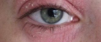
The photo of xanthelasma of the eyelids shows that it can be of small size. It resembles a plaque that forms in the upper eyelid area. Most often, the formation is located closer to the inner corner of the patient's eye.
The plaque protrudes slightly above the surface of the skin, making it visible to the naked eye.
The tumor differs in color from the surrounding skin - it has a yellowish tint. The formation has a soft consistency; when pressed, the patient does not feel pain.
Xanthelasmas can be single or multiple. The tumors usually affect the eyelids of both eyes.
Multiple xanthelasmas can merge into single plaques that have fuzzy edges and a bumpy surface. Often the neoplasms form a continuous thin line that runs along the entire surface of the eyelid.
The formations appear suddenly, without any redness or other changes in the skin of the eyelids. They grow very slowly and gradually spread over the entire surface of the eyelid.
Xanthelasmas usually grow to the size of a small pea; in rare cases, they grow to the size of a bean (approximately 4-5 cm). Tumors do not cause any unpleasant sensations to the patient, with the exception of psychological discomfort from their appearance.
However, tumors significantly affect the patient’s appearance and prevent them from feeling healthy, so they are considered a rather large cosmetic problem.
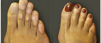
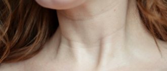
In addition to tumors on the eyelids, patients often develop tumors on other parts of the body - face, neck, limbs, joint surfaces.
Such formations are called xanthomas. Their appearance indicates that the patient has a specific disease - xanthomatosis.
Causes of deposits
Most often, the causes of fat deposits on the skin of the eyelids are the same as atherosclerotic lesions of blood vessels: congenital or acquired hypercholesterolemia of low and very low density lipoproteins. It develops when:
- hereditary breakdown of genes responsible for the synthesis of lipase enzymes or for the formation of cellular receptors that capture “bad” lipoproteins for further utilization;
- endocrine diseases (thyroid gland with hypothyroidism, diabetes mellitus);
- pathologies of the liver and gall bladder;
- chronic inflammation of the pancreas;
- kidney diseases with impairment of their functions;
- long-term use of certain medications;
- obesity;
- long-term intoxication;
- unhealthy lifestyle (physical inactivity, smoking, drinking);
- frequent stressful situations, lack of sleep, night wakefulness and daytime sleep.
In rare cases, a “wen” on the eyelid is one of the manifestations of a metabolic disease such as xanthomatosis. Pathology is assigned a separate code in the international classification of diseases (ICD 10), in contrast to symptomatic xanthomas. With xanthomatosis, plaques form, the cause of which remains unknown, but it has already been established that elevated cholesterol levels have nothing to do with it. Metabolic disorders occur intracellularly in pathologically altered immune cells, histiocytes. At the same time, the lungs and bones are also affected, in which rough scars grow.
TOP 3 disease risk factors
Risk factors for the development of xanthelasma are:
| Factor | Explanation |
| Female | The disease occurs in men, but much less frequently. |
| Patients over 45 years of age | Against the background of age-related changes, the lipid balance in the body is often disturbed |
| People with chronic diseases | Diabetes mellitus, obesity and other endocrine pathologies |
Patients at risk are recommended to undergo regular lipid profile testing to prevent the occurrence of neoplasms.
Reasons: why do xanthomas form?
The cause of this phenomenon is usually associated with changes in lipid metabolism, systemic or rarely local in nature.
- Xanthomas are common among patients with severe forms of hypercholesterolemia and hypertriglyceridemia, usually on a hereditary (primitive) basis, sometimes appearing large in homozygous individuals; these patients have very high levels of cholesterol and/or triglycerides in the blood .
- Xanthomas may appear less frequently even in patients with primary biliary cirrhosis, pancreatitis , diabetes mellitus , heart failure , and some forms of cancer and inflammatory diseases.
Diagnostics
If a tumor occurs in the eyelid area, the patient is advised to make an appointment with a dermatologist. A specialist determines the presence of xanthelasma based on its appearance - a flat yellowish formation in a characteristic location.
Additionally, during the examination, the dermatologist uses the diascopy technique. The formation is compressed with a transparent glass slide. This can achieve bleeding of xanthelasma. Due to the outflow of blood, the yellow color of the tumor is more clearly visible.
It is necessary to differentiate xanthelasma with the following diseases:
- malignant neoplasms of the skin;
- elastic pseudoxanthoma;
- syringoma.
Based on the clinical manifestations of the disease, the specialist diagnoses the patient and sends him for a full examination.
After determining xanthelasma, the patient must undergo a blood lipid test. For this purpose, the level of cholesterol, triglycerides and lipoproteins is determined.
Diagnostics allows you to assess the level of risk for the development of such dangerous diseases as atherosclerosis, hypertension and myocardial infarction.
What are xanthomas?
Xanthomas on the skin are plaques or yellowish nodules caused by deposits of macrophages filled with fat on the body. They appear as soft yellow masses with a distinct edge located just below the surface of the skin (see photo below). The diameter of xanthomas can vary from a few millimeters to more than seven centimeters.
Xanthomas are usually so harmless that they are considered mainly as a simple aesthetic disorder; however, because they often represent a manifestation of high blood lipid concentrations or are accompanied by other serious diseases, they are usually associated with a high risk of disease (especially atherosclerotic-based cardiovascular disease) and other types of diseases.
Eliminating the cause of the pathology
Treatment of xanthelasma can be divided into several main stages. Therapy of the disease is a complex treatment that affects not only the tumor itself, but also the factors contributing to its appearance.
A course of specific treatment is necessary to prevent the recurrence of tumors.
An important component of therapy is the influence on the cause of the development of xanthelasma. Thus, in case of diabetes mellitus, the patient’s insulin therapy is adjusted. If the thyroid gland is malfunctioning, hormone replacement therapy, for example, thyroidin, is indicated.
To reduce cholesterol levels, the patient is prescribed a special therapeutic diet. It involves strict limitation of foods containing large amounts of animal fats. They are replaced with vegetable oils.
Preference is given to olive and flaxseed oil, as they are more beneficial than sunflower oil.
More plant foods are introduced into the diet - vegetables, fruits and berries. It is recommended to stop drinking alcohol during the treatment period.
Nicotine consumption prevents the body from restoring normal metabolism, so the patient should try to minimize the number of cigarettes smoked.
To reduce blood cholesterol levels, special lipotropic drugs are used that normalize the fat balance in the body.
The most effective means include:
- Pyricarbate.
- Lipoic acid.
- Clofibrate and its analogs.
- Thioctic acid.
- Cetamifene.
- Esters of polyunsaturated acids.
To reduce cholesterol, herbal preparations that have fewer side effects can also be used.
Drugs with this effect include:
- dandelion root;
- corn silk;
- Birch buds;
- dog-rose fruit;
- immortelle flowers.
Lipotropic drugs have a pronounced choleretic effect - they stimulate the secretion of bile from the liver. Therefore, their use is prohibited if there are any disorders of the biliary tract.
The use of medications must be agreed with your doctor. The patient may have contraindications to the prescription of drugs, which can be diagnosed by a specialist.
Additionally, to normalize metabolism in the body, the administration of vitamins and microelements is required. The use of multivitamin preparations has a restorative effect on the patient’s body, helping to restore normal metabolism.
TOP 5 methods of tumor removal
After treating the underlying pathology, the patient can remove the tumor. Typically, treatment takes place on an outpatient basis, meaning the patient does not need to go to the hospital to have surgery.
The tumor removal procedure can be performed using several methods. Each of them has its own advantages and disadvantages.
Surgical intervention
The operation involves excision of the xanthelasma using a scalpel. The area of its localization is treated with iron sesquichloride, after which sutures are applied to the edges of the surgical wound.
Surgical intervention injures the human skin and leaves a noticeable scar on the eyelid, so it is usually used for large tumors that cannot be removed by other methods.
In other cases, more minimally invasive treatment methods are used. Surgery is often performed when patients have xanthelasmas and drooping upper eyelids.
In this case, along with excision of the tumor, excess skin tissue above the eye is removed.
The average price for removing eyelid xanthelasma ranges from 5,000 to 12,000 depending on the stage of the disease.
Laser removal
The use of a carbon dioxide laser is an effective and safe method of tumor removal. The device allows you to excise only the tumor tissue without damaging the healthy skin around it.
The main advantages of the technique include:
- Complete sterility.
- No mechanical or thermal injury to the skin.
- High precision of tumor removal.
- Rapid bleeding stop.
- Contactless procedure.
Due to its advantages, laser excision produces a minimal cosmetic defect, which allows it to be used for excision of small xanthelasmas.
The price of the procedure ranges from 6-20 thousand rubles.
Electrocoagulation
Electrocoagulation is the destruction of tumor cells using electric current.
At the first stage of exposure, the protruding parts of the neoplasm are removed using tweezers or scissors. After this, electrodes are applied to the base of the tumor, with the help of which the skin is cauterized.
After the procedure, the patient is left with a small crust on the eyelid that has a reddish color. With proper care, the postoperative wound heals in about 10 days.
In the first few months, a reddish spot remains in its place, which then completely disappears.
The main advantage of electrocoagulation is the elimination of possible relapses.
Treatment of the tumor with electric current leads to the fact that all cells of the tumor die. Therefore, xanthelasma cannot re-develop at the surgical site. The price of electrocoagulation is 1200-1400 rubles.
Cryodestruction
Cryodestruction involves the destruction of neoplasm tissue using targeted exposure to ultra-low temperatures. The device supplies liquid nitrogen, which kills tumor cells. Further growth of the tumor stops completely.
At the site of the procedure, a dry scab is formed - a small fragment of necrotic tissue, which disappears after about 10-15 days. In its place, pink skin is formed, indistinguishable in color from healthy tissue.
The cost of the procedure is approximately 4-6 thousand.
Radio wave tumor removal
The use of radio waves to excise xanthelasmas ensures high accuracy of tumor removal. During the action of waves on the skin, heat is generated by a thin electrode.
Thermal exposure leads to the destruction and evaporation of tumor cells. The use of radio wave radiation provides the finest cut, much more accurate than with a scalpel.
Thanks to this, the skin around the tumor is practically not injured, and the cosmetic defect disappears. The average price of the procedure is 600-1500 rubles.
Classification and symptoms
There are different forms of xanthoma:
| Type of xanthoma | Peculiarities | The type of disease they are associated with |
| Xanthomas of the eyelids (xanthelasmas) | Yellowish plaques are located around the eyelids, the most common place where xanthomas can be found. Usually occur after 50 years of age; they are asymptomatic and usually bilateral and symmetrical | Hyperlipidemia types IIa, IIb and IV are not always associated with lipid metabolism disorders |
| Tuberous xanthomas | Nodules of variable size from 5-6 mm to several centimeters, hard, painless, lobular, flat (see photo above). They develop slowly and mainly affect pressure areas such as the elbows, knees and buttocks | Type IIa homozygous hyperlipidemia |
| Tendon, subperiosteal, fascial, aponeurotic xanthomas | Subcutaneous nodules adhering to tendons, especially Achilles, but also to ligaments, muscle bands and periosteum, mainly of the lower extremities | Type IIa hyperlipidemia |
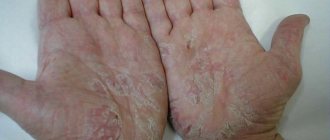
| Flat xanthomas | They appear as spots or thin palpable yellow-orange plates, asymptomatic, of variable size. They can be located in the folds of the face and upper torso; when they occur on the palm, they become a pathognomonic characteristic of dysbetalipoproteinemia type III | Type IIa homozygous and type III hyperlipidemia |
| Eruptive xanthomas | They appear as a sudden eruption of yellow-red papules located on the extensor surfaces of the limbs and on the buttocks. They increase and decrease in size in parallel with changes in plasma triglycerides and lipids | Diabetes and hyperlipidemia types I, IV and V |
Question answer
An important quality of xanthelasma is that the neoplasm never degenerates into cancer.
Therefore, the risk to the patient’s health is practically minimal. Very rarely, patients experience bleeding at the extraction site, and an unsightly scar may also form. In addition, such a plaque can form again. For everything to go well, you need to go to a good specialist.
There are certain rules of prevention. To do this, doctors recommend adhering to proper nutrition, reducing the consumption of carbohydrates and animal fats. It is worth treating diseases that provoke tumor formation in a timely manner, not injuring the eyelids, removing makeup, using good cosmetics, and regularly seeing a dermatologist.
TOP 5 medicinal mixtures (folk methods)
To treat xanthelasma of the eyelid with folk remedies, compresses are often used:
- honey - a mixture of an egg, a spoonful of flour and a spoonful of honey;
- onion – soft onion baked in the oven, mixed with laundry soap;
- sour cream – sour cream in equal proportions with honey;
- chestnut – 5 crushed baked chestnuts with the addition of crushed aloe leaf;
- wheat – ground wheat grains diluted with olive oil.
The prepared mixture must be regularly applied to the surface of the tumor, left for 15-20 minutes, and then washed off with water.
Treatment of xanthelasma of the eyelids with folk remedies using compresses has excellent reviews among patients.
To normalize cholesterol levels in the body, you can use propolis tincture. It must be diluted in purified water, 7 drops per 30 ml. The resulting solution must be taken once every day during lunch.
To treat xanthelasma of the eyelids with folk remedies, you can use herbs. Patients need to brew yarrow, mint or chamomile. They need to be drunk twice a day, 100-150 ml. The effect of the procedure is achieved in approximately 1-2 months.
Patient reviews
The cost of eyelid xanthelasma removal can vary significantly, so it is recommended to read patient reviews before scheduling the procedure.
LARISSA, MOSCOW:
“I had a small plaque in the upper eyelid area, so I decided to remove it with a laser. The procedure was done very quickly, it didn’t hurt me, of course, there were some unpleasant sensations. Everything healed quickly, there were no traces left.”
ALEXEY, ST. PETERSBURG:
“I took my mother for treatment - some kind of yellow tumor appeared on her eyelid.
They said that it was already too big and could only be removed surgically. The operation was done quickly, under local anesthesia. We are pleased with the result, although there is a small scar left on the upper eyelid.”
Expert opinion
- Cosmetologist
- Surgeon
Anna Avaliani
practicing cosmetologist
I think that it is better not to self-medicate using traditional methods, but to go to the doctor. To be sure to get rid of the plaque, it is better to resort to radical excision by any method (laser, liquid nitrogen, etc.). These are minimally invasive techniques, as they do not require the use of general anesthesia.
Aisha Baron
plastic surgeon
You can get confused in the variety of xanthelasma removal methods.
But the most effective way can be considered coagulation using a laser. The procedure has many advantages. After it, rehabilitation does not last long, the plaque will not appear again, the session itself is painless, and the risk of complications is reduced to zero. The procedure will take about 15-30 minutes. Xanthelasmas are neoplasms that are an important prognostic sign. Their occurrence indicates that the patient’s body has a disturbance in the metabolism of fat molecules.
Therefore, you should not delay treatment of the disease. You need to consult a specialist and undergo an examination to discover the cause of the formation of tumors.
Is it possible to cure eyelid xanthelasma on your own without surgery?
To diagnose the disease, no special procedures are prescribed. Plaques are visible to the naked eye. But the ophthalmologist can refer the patient to an endocrinologist and dermatologist to identify problems associated with fat metabolism. Xanthelasmas can only be removed surgically. It is impossible to get rid of them at home using folk remedies or medications. Today, removal of formations on the eyelids is carried out using various methods:
- Classic surgery with a scalpel. The plaques are excised and separated from the eyelid.
- Cryodestruction - cauterization of xanthelasmas with liquid nitrogen.
- A laser procedure during which tumor tissue is evaporated by a laser.
- Chemical destruction - treating the skin with trichloroacetic acid.
- Electrocoagulation is the destruction of plaque tissue with high-frequency current.
Folk remedies for treatment will not be beneficial, but they can be used after surgery. The patient can make lotions from various herbs to soothe the skin, relieve itching and speed up tissue healing.