The appearance of temporary slight discomfort and some passing adverse reactions of the dermis to contouring procedures is absolutely normal.
However, after injections of fillers, atypical compactions of connective tissue often form , in other words, fibrosis . In some cases, you need to immediately consult a cosmetologist.
What tissue fibrosis is, the causes of its occurrence, treatment methods, as well as many other issues, we will consider further.
What happens on the first day after hyaluronic acid injections if necrosis begins
The problem with impending necrosis of the skin and subcutaneous tissue after intra-arterial injection of cross-linked hyaluronic acid is that symptoms do not always appear immediately. Therefore, the patient, satisfied with his appearance, calmly goes home, not knowing about the problem. Then, warned of a recovery period in which swelling, pain and bruising are possible, he ignores the dangerous changes.
It also happens that the symptoms of complications appear immediately, but the cosmetologist, who did not study to become a doctor, ignores the problem.
Possible symptoms of incipient necrosis, appearing after a few minutes or several hours:
- whitening of the skin;
- pain at the injection site;
- venous stagnation.
The pain gradually gets worse despite taking over-the-counter pain relievers. Discoloration of the skin in the treated area becomes stable. The area where hyaluronic acid was injected feels numb.
On the first day, the skin becomes ischemic, compensatory blood flow increases through other vessels, the body tries to protect itself from blood supply disturbances. There is no necrosis yet, but you need to act very quickly.
General concept of fibrous formation
Despite the fact that fillers based on hyaluronic acid are completely compatible with the human body in terms of biological characteristics, nevertheless, after administration of the drug, inflammation may develop, which usually goes away after 2-3 days.
If the formations persist for two weeks or more, you should consult a doctor - probably the inflammatory process has become chronic and the growth of connective tissue begins to be excessive. Such local compaction (the medical term is fibrosis) in the area where the special gel was introduced indicates the beginning of the process of producing new collagen fibers. Capsules of the fibrous type become noticeable not only upon palpation, but also visually (the relief of the skin changes).
Fibrous tissue deformation is considered a late and difficult-to-treat consequence of correction and modeling of facial contours with fillers.
How can you cope with tissue necrosis caused by blockage of a vessel with hyaluronic acid?
Time plays an important role here, and the only causative treatment can only be the introduction of hyaluronidase, which dissolves hyaluronic acid in the vessel. The later this happens, the more irreversible changes will occur in the skin and subcutaneous tissue. Possible complications include extensive scarring and facial artery occlusion. Irreversible changes also affect muscles.
The introduction of hyaluronidase always makes sense, but the later the patient presents, the less the consequences are limited to ischemia. Immediate dissolution of the acid will not leave any permanent marks on the face and the patient will not require additional treatment. If the drug is administered after three days, the acquired pathologies will be visible for many years, if not a lifetime.
In addition to hyaluronidase, the treating physician may also administer an antiplatelet agent, an anticoagulant, an antibiotic, an antiviral drug, etc.
Possible causes
Multiple growing fibrous capsules, lumps and other compactions are the most common complications after filler injection. Of course, only a doctor can determine the cause of their appearance, while in some cases the cause of fibrosis is never established and is considered an individual reaction of the body, but some factors can be listed :
- direct transfer of funds or its low quality;
- incorrectly chosen injection technique;
- development of a focus of inflammation;
- mechanical damage to the walls of blood vessels;
- individual reaction of rejection of a foreign substance by the body;
- in the injection area there is a noticeable growth of connective tissue - scars, nodules or cysts (for example, hyaluronic acid enhances the production of fibroblasts);
- failure to comply with hygiene and care rules, and as a result, infection;
- unprofessionalism of the cosmetologist.
In addition, the formation of fibrous capsules can be provoked by an incorrectly calculated dose of the drug, as well as its too superficial administration.
Differential diagnosis of granulomas after fillers
Sometimes it is difficult to differentiate between granulomas , nodules that form for other reasons, and abscesses. Thus, nodules may appear after contouring if the drug was administered using the wrong injection technique, in case of infection or the development of a late allergic reaction.
Granulomas should be differentiated from nodules that form as a result of improper injection technique, infection or late allergic reaction.
Tests that can help make a diagnosis:
- C-reactive protein;
- number of leukocytes;
- erythrocyte sedimentation rate;
- microscopic examination and culture;
- in situ hybridization;
- CT scan;
- Magnetic resonance imaging;
- biopsy and histological examination of the skin.
Symptoms of epiretinal fibrosis of the eye
The clinical picture of the disease directly depends on the neglect and degree of progression of the process. Epiretinal fibrosis is often an incidental diagnostic finding and is asymptomatic. In such cases, the pathological process proceeds very slowly and does not cause significant visual discomfort. Patients have normal or close to normal visual acuity.
But sometimes active proliferation occurs, and the contractile properties of the resulting connective tissue membrane lead to damage and “curvature” of the retinal surface. With this progression of the disease, the following clinical manifestations occur:
- A common occurrence is metamorphopsia, a phenomenon in which straight objects with clear outlines (for example, a window frame, tile joints or door frame) become curved or convoluted. The presence of distortions and broken straight lines is easy to notice when comparing a healthy eye with a diseased one.
- Objects in the field of view of patients with epiretinal fibrosis may appear larger or smaller than their actual size.
- In advanced cases, banal reading becomes difficult for patients. More rare symptoms are double vision, increased sensitivity to light, blurred vision.
- As the pathology progresses, complete loss of central vision is possible.
| Metamorphopsia | Blurred vision |
The pathological process has a particularly negative effect on the macular region of the retina, leading to disturbances in central vision. Visual discomfort is determined by the degree of deformation, localization of fibrosis and secondary changes in the retina. A significant decrease in visual acuity, as well as the appearance of dark spots in the visual field, is usually associated with the formation of traction macular edema, retinal tears, traction maculoschisis, or even retinal detachment.
Diagnosis of macular epiretinal fibrosis
The specific and simplest diagnostic test for this pathology is the use of the Amsler grid. The grid itself is a square lined grid measuring 40 by 40 cm. There is a dot in its center. The patient should look at the grid image without interruption with each eye in turn. The wrinkling of the retina caused by epiretinal fibrosis causes the lines to become curved and appear wavy. This is the simplest screening test. You can do it at home by first downloading from the Internet and printing a picture with an Amsler grid.
The simplest method is ophthalmoscopy - epiretinal fibrosis can be easily visualized on the surface of the retina in the form of a translucent whitish film, which has a characteristic folding, shines and resembles oilcloth in appearance.
Another diagnostic option is an ultrasound examination of the eye, which allows one to visualize gross fibrotic manifestations of an advanced stage of the disease. However, at the initial stages of pathology, such a diagnostic test may be uninformative.
Fluorescein angiography is not important for making a primary diagnosis. To a greater extent, it allows us to identify complications associated with the presence of epiretinal fibrosis. In advanced cases, it visualizes tortuosity of retinal vessels or macular edema. Fluorescein angiography can detect retinal ischemia, which significantly affects the preoperative prognosis of visual function after removal of epiretinal fibrosis.
| Ophthalmoscopic picture | Fluorescein angiography |
Optical coherence tomography (OCT) helps to most reliably determine the presence or absence of macular epiretinal fibrosis. This research method allows us to objectively assess the dynamics of development and the effect of fibrosis on the condition of the retina. Namely, to confirm the presence of its thickening, macular edema, as well as any traction effects on the retina from the vitreous body. Using OCT, ophthalmologists also monitor the course of the postoperative period.
Case from practice: how lips turned into dumplings. Complications with lip augmentation
For some reason, it is generally accepted that natural preparations such as hyaluronic acid cannot cause complications and allergies. This is an absolute mistake. After all, there are allergies to natural products - strawberries, oranges and even cabbage, and this does not surprise anyone.
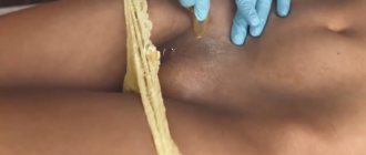
Recently a girl came to our Center. She had her lips enlarged and within a week of the procedure she began to experience swelling. The patient went through a couple of specialists, who told her that everything would pass soon. But the swelling did not go away, and her lips began to hurt, so the patient signed up for an aesthetic medicine clinic - with a certified dermatologist-cosmetologist.
Our doctor noted monstrous swelling in the lip area and inflammation, visible to the naked eye. The girl’s lips hardened so much that it became difficult for her to speak. Treatment involved removing hyaluronic acid and treating allergies and inflammation with anti-inflammatory drugs and steroids.
Reaction to lip augmentation with hyaluronic acid
Everything took more than 20 days. The lips, fortunately, recovered - the inflammation stopped, the acid dissolved, and the mouth returned to its original shape.
The worst thing in this situation is that the patient visited the doctors, and no one helped her. Cosmetologists, counting on the safety of hyaluronic acid, underestimated its case, showing complete ignorance in this matter. But the treatment in this case was quite simple.
Clinical and histological features of granulomas after contour plastic surgery
Depending on the histological properties, there are four types of granulomas after dermal fillers:
- cystic (HA, bovine collagen);
- nodular lipogranulomas (silicone, polyacrylamide);
- sclerosing;
- mixed.
| Intralesional injections | Systemic therapy | Surgical methods |
| Corticosteroids (triamcinolone acitate – Kenalog) Bleomycin 5-fluorouracil | Antibiotics Prednisolone Allopurinol Colchicine Cyclosporine | Laser removal Opening and drainage Excision and fat transfer or flap |
Rice. 2: granuloma treatment methods
Clinically, granulomas appear as red, firm papules, nodules, or plaques that may appear months or years after filler injections.
Consequences and measures to prevent the situation from getting worse
The appearance of fibrous nodes after the filler injection procedure is not a fatal condition. Fibrosis is usually perceived as a kind of cosmetic defect of the skin. However, mechanical damage to such a seal may result in complications:
- minor bleeding;
- wound infection;
- pain syndrome;
- necrosis of injured tissues.
The last of the listed complications - tissue necrosis - is worth considering in more detail, due to the fact that it is the most undesirable, unpleasant of all possible consequences of the injection of fillers. In addition, the result of such a complication can even be fatal.
What is epiretinal fibrosis?
Epiretinal fibrosis of the eye consists of the formation on the inner surface of the central part of the retina of a transparent film or membrane consisting of connective tissue cells. Such connective tissue membranes have contractile properties, that is, they can gradually contract with the formation of retinal folds, deformation of the correct contour of the retina, which contributes to progressive deterioration of vision and pronounced metamorphopsia due to the traction effect exerted on the underlying layers of the retina.
There is currently no single theoretical basis explaining the causes and mechanisms of development of epiretinal fibrosis. According to the generally accepted theory of Roth AM and Foos RY, the main trigger for the development of this disease is partial vitreous detachment with its pathological adhesion to the macular zone of the retina.
According to recent research data, about 70% of all cases of epiretinal fibrosis are associated with posterior vitreous detachment. More often, this condition is age-related, develops without concomitant eye diseases and is called idiopathic epiretinal fibrosis. However, epiretinal fibrosis of the eye can also be present in various eye diseases and pathological conditions of the organ of vision. In this connection, in the practice of ophthalmic surgeons, the term secondary epiretinal fibrosis was adopted, that is, associated with other ophthalmological conditions:
- Diabetic retinopathy.
- Hemorrhages into the vitreous cavity.
- Inflammatory diseases of the eye (iridocyclitis, uveitis, panuveitis).
- Thrombosis of the retinal vein or its branches.
- Retinal tear or detachment.
- Injuries and injuries to the eye.
- History of ophthalmic surgery.
| Dense connective tissue membranes on the surface of the retina |
Statistically, the risk of epiretinal retinal fibrosis increases with age. Studies have shown that this pathology manifests itself in 2% of patients over 50 years of age. For people over 75 years old, this figure is already 20%. Both men and women are equally susceptible to the disease. In 10-20% of cases, bilateral damage occurs, that is, epiretinal fibrosis develops in two eyes, but the severity of the manifestations may vary.