The mammary glands in boys and girls are identical before puberty. At the age of 12-13, the male body begins to synthesize the hormone testosterone, which should be used to build a male body and form secondary sexual characteristics. Where then does enlarged breasts come from - gynecomastia in adolescents?
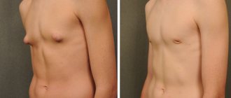
The fact is that many boys enter adolescence overweight. Malnutrition - lack of a full breakfast and frequent consumption of fatty and sweet foods leads to excess deposition of fatty tissue in the hips, lower back, abdomen and chest. This is also facilitated by the refusal of sports activities and a sedentary lifestyle. As soon as puberty occurs, the male hormone testosterone can be converted in excess into the female sex hormone estradiol, which forms breasts in such a teenager according to the female type and develops gynecomastia.
There is another option - physiological gynecomastia, associated with a sharp increase in testosterone levels, which goes away on its own in adolescents within a short time after its onset.
Classification of gynecomastia
There are two types of gynecomastia: false and true.
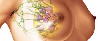
False is not a real disease, but only the deposition of adipose tissue in the chest area. With true, pathological growth of glandular tissue occurs.
True gynecomastia can be of the following types:
- Idiopathic
- Physiological (neonatal, pubertal, senile)
- Pathological
- congenital (with Klinefelter's Syndrome, anorchia, hermaphroditism, androgen resistance syndrome, enzymatic defects in testosterone synthesis, increased aromatase in peripheral tissues)
- endocrine (for castration, mumps, Cushing's syndrome, congenital adrenal hyperplasia, adrenocorticotropic hormone deficiency, hyperthyroidism, hypothyroidism, panhypopituitarism, hyperprolactinemia)
- symptomatic (for tumors of the testicles; adrenal glands; pituitary adenoma; breast cancer; tumors that secrete human chorionic gonadotropin)
- medicinal (when using hormones; antiandrogens; prolactin stimulants; when using drugs; anti-tuberculosis drugs)
- metabolic (with thyrotoxicosis; renal failure; liver cirrhosis; fasting; alcoholism)
- other (for HIV, chest wall trauma, cystic fibrosis, physiological stress)
Diagnostics
A thorough medical history is important to determine the underlying cause of gynecomastia and rule out breast cancer and other tumors. On the other hand, it should be noted that about 25% of cases of gynecomastia may be idiopathic (the cause is unknown).
Must be taken into account during examination
- patient's age,
- onset and duration of breast augmentation,
- symptoms of associated pain,
- recent weight change and
- possible endocrine disorders.
It is necessary to establish whether any medications or drugs are being taken, as they can cause up to 10–20% of cases of gynecomastia.
On physical examination, gynecomastia is defined as overgrown glandular tissue under the nipple-areolar complex and can vary in size.
True gynecomastia must be differentiated from pseudogynecomastia, or false gynecomastia, which is manifested by hypertrophy of adipose tissue without proliferation of the gland itself.
Breast cancer is rare and accounts for less than 1% of malignant neoplasms in men (according to UK and US data). It usually appears on one side and is felt as a hard mass located outside the nipple-areolar complex.
As part of a systematic review, the physical examination should include a search for signs of chronic disease of the thyroid gland, kidneys or liver, or genital organs. When examining the genitals, it is also important to monitor testicular weight or testicular atrophy. Sometimes liver enlargement may occur.
Laboratory research
Biochemical evaluation includes tests to determine liver, kidney and thyroid function, as well as serum levels of testosterone, prolactin, follicle-stimulating hormone and luteinizing hormone.
Additional tests may be necessary in cases of recent or symptomatic gynecomastia to rule out tumors. For example, serum levels of estrogen, human chorionic gonadotropin (hCG), dehydroepiandrosterone and 17-ketosteroids in urine.
Routine mammography and breast ultrasound with or without biopsy are usually not recommended unless breast cancer is suspected or the patient has unilateral breast enlargement.
Similarly, a testicular ultrasound or CT scan of the abdomen may be performed if the testicle or adrenal gland becomes enlarged.
Diagnostic evaluation of gynecomastia
- Lab tests: testosterone
- estrogen-β-human chorionic gonadotropin
- prolactin, follicle-stimulating hormone and luteinizing hormone
- urea and electrolytes
- ± dehydroepiandrosterone or 17-ketosteroids in urine
- Ultrasound of the breast
Patient selection
Various clinical and histological classifications have been proposed for gynecomastia. The Simon classification is the most practical because it takes into account not only breast size, but also the amount of excess skin.
Diagnostic features
The doctor determines the presence of pathology through visual examination of the mammary glands and palpation. The boy's genitals can also be examined. Also mandatory studies for suspected gynecomastia are:
- Ultrasound of testicles and mammary glands;
- analysis of the patient’s blood for the content of hormones;
- X-rays of light;
- mammography, breast biopsy if there are lumps in it.
Only as a result of a full examination will the doctor be able to make the correct diagnosis and determine the direction of further treatment.
Indications for removal of gynecomastia
- Persistent increase after puberty (>2 years)
- Inadequate response to treatment
- Severe breast enlargement
- Significant asymmetry or unilateral condition
- Patient request
- Post-massive weight loss
- Special clinical conditions
The boundaries between categories are not clearly defined, which leads to subjectivity. Therefore, in our practice, we classify gynecomastia into three stages:
- small to moderate in size with no or minimal excess skin (Grades I and IIA)
- medium to large in size with moderate to severe excess skin (grades IIB and III).
Exercises
At the early stage of gynecomastia, the production of male hormones is activated with the help of physical activity. In addition, regular activity helps stabilize weight, this is important when the mammary gland increases in size.
At home, regular push-ups will be useful, removing fat cells in the problem area and helping to build muscles in the upper extremities. Raising and bringing your arms together with dumbbells in a lying position will help bring the mammary glands into proper shape.
For simple exercises to be beneficial, you need to take classes under the guidance of a trainer. He will develop an individual set of classes designed for a specific teenager with existing health problems.
Treatment of gynecomastia
Drug treatment
Most cases of gynecomastia do not require treatment as they are benign and self-limiting.
Weight loss should be recommended first for patients with pseudonecomastia. Particular attention should be directed to eliminating any identified cause.
Drug therapy is mainly aimed at correcting androgen and estrogen imbalance. Medicines are most effective during the active, proliferative phase of gynecomastia. In patients with prolonged gynecomastia for more than 1 year, drug treatment is often ineffective, since the breast tissue progresses to irreversible dense fibrosis and hyalinization during this time.
Treatment of gynecomastia without surgery involves taking medications. Antiestrogenic drugs are often prescribed.
The most common drugs:
- Tamoxifen
- Clomiphene citrate
- Proviron
- Progestogel.
Taking medications must be agreed with your doctor. It is he who prescribes the treatment regimen, determines the required dosage and duration of medication. You should not self-medicate, as this can greatly worsen the patient’s health.
Surgery
The mainstay of treatment for gynecomastia is surgical treatment, the goal of which is to restore the normal-appearing contour of the male breast with minimal scarring while maintaining the viability of the nipple-areolar complex.
Surgical treatments for gynecomastia include various forms of liposuction, breast excision, skin tightening, and a combination of these.
Liposuction is now a major surgical technique because it is minimally invasive, improves contouring by smoothing the contours, and is often successful as a stand-alone technique.
Various types of liposuction have been described, including conventional, mechanical, ultrasound, laser and vibration assisted (VASER). The most common types are conventional and ultrasonic liposuction.
Anesthesia and infiltration
The operation is performed under general anesthesia. All patients undergo preoperative photography. All patients receive antibiotic prophylaxis.
Patients are marked before surgery in a vertical position, indicating the inframammary fold, the boundaries of the mammary glands, and the planned incision sites.
The breast tissue is infiltrated through an incision in the inframammary fold, and a special raster for liposuction is introduced.
Liposuction allows you to achieve good breast contours with minimal scarring. Special cannulas for liposuction are used.
Open gland removal
Open removal of gland tissue is performed through the lower areolar approach. Various other incisions are also described, such as circumvertical, periareolar, transareolar, anchor, t-inverted.
Liposuction is often not effective for removing dense glandular tissue. Excisional techniques are effective for these patient populations, although they are associated with a high complication rate with the potential for gross scarring and contour deformities.
We use open excision combined with liposuction. Liposuction serves several purposes, such as pretunneling to facilitate resection, reduce bleeding and bruising, and partially destroy breast tissue. After liposuction, tissue can be removed through several small incisions.
We prefer the time-tested method of open excision, where the breast tissue is excised through a semicircular incision along the lower edge of the nipple-areolar complex. At least 1 cm of breast tissue is left under the areola to prevent depression of the nipple-areolar complex.
Open excision, even in combination with liposuction, sometimes does not completely correct large and ptotic breasts for which skin tightening is indicated. Some surgeons recommend avoiding skin resection, allowing the skin to shrink on its own for 6-8 months. We prefer to remove excess skin during the first operation. This has practical advantages such as reduced cost, single operation, and reduced operating time.
Skin tightening
There are a number of techniques used to tighten the skin of gynecomastia. Although most of them are similar to those used for mastopexy in women, there are still some differences.
Although skin tightening techniques may result in larger scars compared to liposuction and simple removal, they are indicated for patients with large breasts, significant ptosis, or poor skin turgor. Therefore, in patients with obvious excess skin or very large breasts, skin tightening techniques should also be planned in advance and should be performed simultaneously with open breast tissue removal or in a second stage, at least 4-6 months later.
However, there is no consensus on when and how to perform skin tightening. We base this decision on skin elasticity, presence of ptosis and excess skin, skin type, and the patient's willingness to accept a potential two-step procedure. Surgeon preference also plays a role in the choice of technique. Whenever there is excess skin or ptosis, we prefer to address the excess skin at initial surgery, especially in cases of poor or borderline skin elasticity.
The choice of skin tightening method also largely depends on the surgeon's preference. It is possible to perform a lift
- circular
- vertical
- without vertical scar with transposition of the nipple-areolar complex
- anchor
Periareolar mastopexy
Periareolar skin tightening is a preferred method over other skin tightening methods, mainly because it avoids additional scarring.
We prefer to use the periareolar technique due to less noticeable scarring.
Vertical mastopexy according to Le Jour
In patients with true ptosis or very large breasts, skin tightening and tissue resection can be undertaken using the LeJour vertical lift. The skin and underlying breast tissue are resected in a vertical ellipse, and then the wound edges are approximated.
However, a vertical scar may be noticeable. This technique is used as a last resort, especially in those patients who want to avoid a T-scar.
Other skin tightening methods
Lateral wedge, elliptical and inverted T-shape excision are sometimes used.
Patients with very large or ptotic breasts are suitable candidates for elliptical mastectomy and free nipple transfer.
This also avoids the characteristic scarring associated with mastopexy in women.
In patients at high risk of developing keloids or hypertrophic scarring, we often use a non-vertical scar technique with Lalonde breast reduction. After deepithelialization, liposuction is performed before open removal of the glands and through a horizontal incision. Once hemostasis is achieved, the nipple areolar complex is perfectly transposed and brought into a new position.
Gradual skin reduction
Patients with mild ptosis sometimes do not need to undergo gland removal with simultaneous skin tightening. The operation can be performed in two stages. We start with liposuction followed by skin tightening after at least 4-6 months.
Sometimes patients scheduled for two-stage surgery are satisfied with the initial results and refuse a second skin tightening procedure because they can experience gradual skin tightening on their own.
However, the skin may still not shrink, in which case we perform one-stage skin tightening and excisional techniques for severe gynecomastia with significant excess after liposuction.
Recovery after gynecomastia removal
Drains are not usually used except in major resections or when skin resection is performed, such as in patients with massive weight loss. Patients are advised to wear a special compression bandage for gynecomastia day and night for 4-6 weeks.
Oral antibiotics are taken for a total of 5 days. Painkillers should be used, as a rule, for 2-3 days.
Outcomes, prognosis and complications
Only an integrated approach can lead to predictable results in the surgical treatment of gynecomastia. Our starting point for all cases of gynecomastia is liposuction, even in those with hard subareolar glands, as it facilitates subsequent open excision. It is also technically easier to perform liposuction at the beginning of the operation rather than after removal. We use conventional liposuction only when ultrasonic liposuction is not available.
In patients in whom open excision is not necessary due to appearance and consistency, we always agree to open excision if liposuction leaves significant residual stromal tissue.
Whenever there is excess skin and/or ptosis, we prefer to treat the excess skin with primary surgery, especially in cases of poor or borderline skin elasticity. This is done to avoid post-operative excess skin.
The choice of skin reduction technique largely depends on the surgeon's preference.
Possible consequences of gynecomastia
- Bleeding and hematoma formation
- Seroma formation
- Infection
- Skin/nipple necrosis
- Contour deformity such as nipple inversion or depression
- Altered nipple sensation
- Redundancy of skin
- Residual asymmetry
- Unfavorable scars (wide, hypertrophy, keloid, pigmentation)
- Overcorrection, undercorrection
- Occasional need for revision surgery: asymmetry, residual tissue, depressed nipples
Difficulty of choice
The method by which gynecomastia is removed is always determined by the surgeon, taking into account the stage of the disease and the individual characteristics of the patient, the technical capabilities of the clinic and personal preferences in choosing a particular technique. Often the question of choice depends on the financial aspect of treatment and the level of equipment of the hospital.
The surgeon determining the tactics is based on certain principles:
- Any operation must end with excision of glandular or adipose tissue;
- The laser injures tissue less, but does not always act radically;
- The incision with a classic mastectomy is larger and the scar is more noticeable, but this circumstance should not be decisive when choosing a technique;
- Control of the volume of tissue excised during conventional surgery is much better than during laser treatment;
- Laser removal is safe only in the absence of symptoms of malignancy, while in case of signs of cancerous transformation, mixed and false varieties of the disease, 3rd degree enlargement of the mammary glands, the open method should be chosen as the safest.
In recent years, due to the widespread introduction of laparoscopy in various fields of surgery, minimally invasive endoscopic techniques have been proposed instead of traditional mastectomy. The essence of laparoscopy for gynecomastia in men is to perform the operation through a small puncture in the armpit area, which ensures the absence of scars later.
Endoscopic treatment of gynecomastia is labor-intensive, requires high professionalism of the surgeon and the availability of expensive equipment, so it is not available in all surgical clinics. In addition, it cannot be used if changes in the gland are likely to be malignant.
Recurrence of gynecomastia
Patients with gynecomastia in general are a surgically challenging group of patients, not least because of their high expectations from surgery. In patients who have undergone surgical treatment, it is sometimes necessary to undergo revision surgery several months after the initial surgery for various reasons. These include inadequate correction, patient dissatisfaction, painful residual lumps, asymmetry, and recurrence (often associated with weight gain). Overcorrection is rare.
Recommendations
It is often difficult for even experienced surgeons to choose the most appropriate treatment. Sometimes a combination of these methods is necessary. Surgical treatment of gynecomastia consists of three main stages, which may not all be necessary for a given patient: liposuction, open excision and skin tightening.
Liposuction should always be used for diffuse or large breast enlargement. It may not be necessary when correcting small breasts with a hard subareolar nodule, but facilitates subsequent open removal. Our results with ultrasonic liposuction have led us to the conclusion that, when available, it is preferable to conventional liposuction because it is more effective and stimulates better skin contraction. Additionally, there may also be less bruising. After liposuction, the consistency of the breast is again assessed and open excision is performed if residual gland tissue is present.
Skin tightening is indicated if there is noticeable excess skin after removal. The choice between a concentric, vertical scar (LeJour technique) and an elliptical skin excision largely depends on the amount of excess skin to be removed, as well as the experience of the surgeon.
Symptoms and signs of pathology in different age categories
The disease manifests itself as a list of defining symptoms that can be used to identify the presence of a pathological process in the patient. In addition to enlargement of the mammary glands, when their volume can reach 10 and sometimes 15 cm, and the volume varies within 150 grams, the disease also manifests itself in other phenomena:
- In the area of the glands there are painful compactions of a homogeneous nature.
- Darkening of the areolas and nipples occurs, and excess pigmentation is acquired.
- The areolas increase in diameter, expand, and develop according to the female type.
- In some cases, the nipple can retract and become flat.
- The boy may complain of pain and heaviness in the chest.
- There may be clear or whitish discharge from the nipples.
In addition to the above, there are other signs, if present, you should immediately consult a doctor and get immediate treatment. These are unbearable pain in the chest, bloody discharge from the nipples, the formation of cracks in the areolas, damage to the skin, an increase in the volume of the axillary lymph nodes, and the formation of papules on the chest. Such signs may indicate the beginning of the development of carcinoma - a malignant tumor, which is extremely dangerous, so you need to consult a doctor immediately!