What is atheroma on the face
Atheroma is a cyst-like tumor under the skin that appears due to blockage of the sebaceous gland duct. It can occur on any part of the body except the palms and feet.
Atheroma is a cyst-shaped tumor that appears due to blockage of the sebaceous gland duct. Often occurs on the face due to the abundance of sebaceous glands.
Causes: seborrheic dermatitis, hormonal imbalance, hyperhidrosis, metabolic diseases.
Symptoms: painless elastic subcutaneous formations; with suppuration, sharp pain, foul-smelling discharge, and fever appear.
Diagnostics: examination, consultation with a surgeon. Treatment: removal with a scalpel, laser, radio wave methods.
However, more often this occurs in places where a large number of alveolar glands are located, namely in the area of the nasolabial triangle, ears, forehead, eyelids and cheeks. Less often, atheroma forms on the cheekbone.
When a blockage occurs, the gland canal fills with sebaceous fluid and stretches. Subsequently, cavities with contents are formed, which include sebaceous fat, epidermal cells, hairs, and cholesterol crystals. The atheroma is surrounded by a capsule that does not allow the contents to spread to nearby tissues.
It is better to entrust the removal of atheroma on the face to a plastic surgeon.
You can also have the operation performed by an oral and maxillofacial surgeon. It is important to make the scar inconspicuous and aesthetically acceptable.
Dermatovenerologist, cosmetologist
Zhikhoreva Inna Viktorovna
6 years experience
Cysts of the sebaceous glands are multiple - that is, when there are several of them, they are small in size and located next to each other. In this case, the ducts are blocked by cholesterol plaques. This process is called atheromatosis.
The neoplasm is benign and does not pose a threat to life. But treatment of atheroma should still be carried out to prevent complications.
A clogged sebaceous gland has the ability to become inflamed and suppurate, which is dangerous due to the development of an abscess in the affected area. Also, a large formation compresses nearby tissues, disrupting normal blood circulation in them.
The need for treatment and its types
Treatment and removal of atheroma is mandatory. Despite its initially benign nature, it can pose a health threat. It is expressed as follows:
- suppuration;
- the appearance of an unpleasant odor;
- breakthrough of the contents of atheroma and formation of ulcers;
- degeneration of a tumor from benign to malignant.
Treatment of atheroma is carried out by removing it. It is carried out exclusively in a hospital setting; manipulations carried out independently lead to complications and wound infection.
Modern aesthetic medicine practices tumor removal by surgery, laser or radio waves. The laser is considered the most advanced. In its pure form, it is used when the size of the atheroma is less than 5 mm in diameter; in all other cases, complex laser and surgical treatment is practiced.
Reasons for appearance
The causes of atheroma on the face are conventionally divided into external and internal. The former include hot climates and environmentally polluted areas - they contribute to hypersecretion of both sweat and sebaceous glands.
Internal reasons:
- seborrheic dermatitis (oily form);
- presence of acne on the face;
- dysfunction ;
- diseases of the anterior lobe of the pituitary gland;
- hyperhidrosis – excessive secretion of sweat glands;
- skin damage due to bacterial infection;
- hereditary predisposition;
- disruption of metabolic processes in the body;
- diseases of the adrenal cortex;
- reduced immunity.
Men often experience atheroma on the face as a result of increased production of testosterone in the blood. But in women, gland blockage can occur due to the application of oily cosmetics to the skin. Failure to comply with hygiene rules leads to contamination of pores and ducts, which is dangerous due to the formation of atheroma.
According to dermatologists, increased secretion of sebaceous glands on the face mainly occurs in teenage girls. As girls grow older, sebum production decreases in girls, and this happens faster than in men.
Why can our articles be trusted?
We make health information clear, accessible and relevant.
- All articles are checked by practicing doctors.
- We take scientific literature and the latest research as a basis.
- We publish detailed articles that answer all questions.
Women's skin dries out more and more with age, while in young people testosterone levels increase, and, as a result, more secretory fluid is produced, increasing the risk of developing atheromas on the face.
However, the cause of atheroma is not only the intense work of the sebaceous glands, but also their dystrophy. This occurs either during the aging period of the body, or in the presence of certain pathologies.
Possible complications
Possible complications after removal of atheroma:
- bleeding - after surgery, laser methods and radio waves weld blood vessels together after the procedure;
- infection - due to improper care, most often - behind an unhealed suture;
- burn - occurs when a doctor makes a mistake in laser and radio wave methods;
- loose seams;
- relapse - reappearance of atheroma if the membrane was not completely removed or the conditions under which the sebaceous glands were clogged persisted;
- the formation of a rough suture is associated either with insufficient experience of the surgeon, or with the characteristics of the patient’s body.
Symptoms
Atheromas can be congenital or acquired. The former have a dense consistency, are small in size, and do not cause pain. Their main localization is the scalp and scrotum area.
Such cysts appear due to abnormalities in intrauterine development; they are rarely diagnosed on the face. If a small child has a formation on his head, doctors immediately recommend removing it, as there is a risk of thrombosis of the blood vessels in the brain.
An acquired (as well as retention or secondary) sebaceous gland cyst can cause pain, bluish appearance, and a round shape. At the beginning of its development, atheroma is almost invisible, only after a while a person can see a small bulge under the skin in the shape of a ball on his face.
Removal of atheroma
Removal of atheroma is the only way to get rid of a cosmetic defect with minimal likelihood of tumor re-formation.
As the capsule fills with sebaceous secretion, the formation will increase. If measures are not taken to eliminate it, the cyst can reach the size of a chicken egg or become infected without having time to grow.
Suppurating atheroma of the face will be accompanied by symptoms such as:
- sharp pain in the affected area;
- swelling and redness of the skin around the tumor;
- increase in cyst size;
- softening of the tumor due to the presence of pus in it;
- weakness and general malaise;
- increased temperature (does not happen to everyone).
The formation of pus in the capsule occurs due to the fact that its contents are an excellent breeding ground for the proliferation of “bad” bacteria. If the cyst is not surgically opened in time, some of the pus may come out on its own, and the rest of the contents may go under the skin. This will lead to a soft tissue abscess or even cellulitis.
It happens that the inflamed atheroma is aseptic in nature, that is, without the formation of pus in it. This process occurs due to traumatization of the affected area. Even slight friction or squeezing often provokes inflammation of atheroma on the face.
In this case, the disease is much easier and does not require emergency surgical intervention. The symptoms will not be as bright as with suppuration and will not be systemic in nature.
Over time, redness, pain and swelling go away. But around the capsule of the sebaceous gland, dense connective tissue will form, which will cover the cyst with a difficult-to-permeable membrane.
Causes of the disease and characteristic symptoms
Most often, atheromas are caused by obstruction in the sebaceous glands or a violation of the outflow of fat in the skin . The following phenomena provoke such processes:
- injuries;
- swelling and inflammation of hair follicles;
- hyperhidrosis;
- metabolic disease;
- bad weather conditions;
- poor working conditions;
- taking antidepressants.
Cystic formations can appear on such parts of the body as:
- head;
- face;
- area behind the ear;
- brow ridges;
- cheeks;
- neck;
- back;
- sacrum and perineum;
- scrotum or labia.
The consistency of the formations is soft, there is no painful sensation, the skin over the formation does not change color . Such formations are the first stage of atheroma and any inflammation leads to rapid tumor growth. Then the skin becomes bluish or inflamed red, ulcers or breakouts begin to appear with the release of pus.
Associated symptoms with atheroma:
- general weakness;
- malaise;
- a sharp increase in temperature.
The disease most often affects those who eat a lot of fried and fatty foods, have acne on the face , and such people have a history of seborrheic keratoses. If you have the first inflammatory symptoms, you should consult a doctor to get rid of atheroma. If this is not done in time, serious complications may occur. Next, we will talk about the features of atheromas on the head and face.
Diagnosis of the disease
One of the doctor's jobs is to rule out the presence of cancer. To do this, part of the formation (usually after its removal) is sent for histological examination.
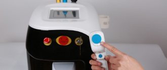
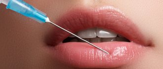
Removal of atheroma using radio waves
Radio waves are a method for removing painless atheromas, the size of which does not exceed 3 centimeters.
When examining a patient, the doctor will easily identify facial atheroma by its external signs:
- visualization of clear boundaries of education;
- dense, elastic fabric structure
- relative immobility of the cyst upon palpation;
- in the central part of the surface of the atheroma there is a clogged duct , which is highlighted in black or bluish color.
Atheroma is usually differentiated from a lipoma, since they are similar in appearance. Some people call the formation of a sebaceous gland a wen, although this is incorrect.
During palpation, the surgeon can easily distinguish atheroma from lipoma: in the first case, when pressing with a finger on the bulge, it remains in place and does not change its shape.
In the second case, education seems to escape from under the fingers in the other direction. Thus, it is impossible to press it. The consistency of lipoma is distinguished by its softness and plasticity.
When excising a tumor, the doctor will immediately notice its membrane. By cutting the capsule in half, its contents will be clearly visible in the form of a curdled mass and you will be able to smell a peculiar unpleasant odor.
Atheroma of the eye area
If the atheroma is located near the eyelid, it will look like a small white bump. It is often confused with a lipoma, papilloma, senile wart or chalazion. Atheromas on the upper eyelid are twice as common as on the lower eyelid, since there are more sebaceous glands in this area.
During night sleep, the secretory fluid, together with the tear fluid, flows into the medial corner of the eye and collects there, so in the morning the formation is especially pronounced. Cysts located near the eyes tend to become inflamed.
Nasal atheroma
The largest sebaceous glands are located on the nose. Most often they are observed on the outside of the wings of the nose, especially on the inside, since multiple small hairs grow there. Atheromas of the nasal area are differentiated from inflamed acne, boils on the inside of the nose, and dermoid cysts.
Formations of sebaceous glands, both small and large, can be observed on the forehead and cheeks. In these places, the formation of atheromatous processes is possible.
In the forehead area, the main number of sebaceous glands are located closer to the scalp; accordingly, blockages of the ducts occur there more often. On the cheek, atheromas are less common, but they bring a lot of trouble, since the skin in this area has greater relief.
Operation stages
Laser removal of atheroma is carried out under local anesthesia and takes 1 - 1.5 hours. The operation involves the following mandatory steps:
- antiseptic treatment and pain relief;
- cutting the skin with a laser beam;
- evaporation of a capsule with liquid contents;
- treatment of the remaining cavity, sealing of vessels, and in rare cases, suturing.
Surgery to remove atheroma with a laser
If the atheroma is of a significant size, the operation is performed both surgically and using a laser. In this situation, manipulations are built according to the following algorithm:
- antiseptic skin treatment;
- general anesthesia;
- cutting the skin with a laser beam;
- mechanical removal of the capsule;
- treatment of the cavity with a laser beam;
- suturing.
The rehabilitation period after the procedure is determined by the type of intervention and anesthesia used. Immediately after the operation, a hole is formed at the site of the atheroma, which is completely healed within 2 - 3 weeks.
Removed atheroma
Treatment methods
In the case of facial atheroma, the most appropriate treatment is considered to be only removal. By itself, even with the use of medications, the formation never disappears, it can only decrease.
Self-medication is dangerous with complications!
Attention
Despite the fact that our articles are based on trusted sources and have been tested by practicing doctors, the same symptoms can be signs of different diseases, and the disease may not proceed according to the textbook.
Pros of seeing a doctor:
- Only a specialist will prescribe suitable medications.
- Recovery will be easier and faster.
- The doctor will monitor the course of the disease and help avoid complications.
find a doctor
Do not try to treat yourself - consult a specialist.
There are several removal methods, everyone can choose the most suitable one for themselves. Conservative treatment is prescribed only to relieve inflammation, if any. After the severe symptoms subside, it is recommended to begin removing the cyst.
When trying to squeeze out atheroma, there is a high risk of causing even more harm to yourself, but you still won’t be able to get rid of it. In the best case, the contents of the gland will come out, after which they will accumulate again and form a mass.
The worst outcome is when, if antiseptic rules are not followed, the wound becomes infected, which will require immediate medical attention.
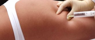
Surgical excision of atheroma on the face
This method of treatment is used for atheromas of any size, provided there is no inflammatory process. If the formation is too large or located near the main vessels, the patient is sent to a hospital for surgery. In other cases, cyst excision is performed on an outpatient basis.
The difference between lipoma and atheroma
New growths on the body, called wen, are similar to various skin diseases.
Surgery involves making a skin incision under local anesthesia and removing the contents of the cyst along with the membrane.
The number of stitches used depends on the size of the wound. A cosmetic suture is usually used on the face, but it will cost more, so not everyone wants to use it. Such nuances should be discussed with the surgeon in advance.
Laser removal
There are three ways of this treatment:
- Photocoagulation – used for non-inflamed and suppurating atheromas of small sizes (up to 0.5 cm in diameter). No stitches are required after the operation, since a protective crust is formed at the site of the removed lesion, under which the wound heals safely in 1–2 weeks.
- Laser excision with sheath - suitable for removing any atheromas measuring from 0.5 to 2 cm. The doctor makes an incision in the skin with a scalpel, then applies laser vaporization near the cyst sheath. At the end of the procedure, a drainage is placed and a suture is applied.
- evaporation of the capsule is intended for removing atheromas larger than 2 cm in diameter. Doctors rarely take on festering atheroma of this size.
Laser treatment is performed under local anesthesia. The postoperative period requires monitoring the wound for the next 10-12 days, wearing sterile dressings.
Radio wave removal
For small formations with no inflammation, doctors can offer a radio wave method for treating atheromas. It is easily tolerated by patients, but is not suitable for everyone.
Treatment involves exposure to radio waves, which provoke cell death only at the site of the formed cyst. After the procedure is completed, a natural crust appears on top, which promotes wound healing.
If the atheroma has festered, it is recommended to open it surgically, clean out the capsule particles from the tissue, rinse with antiseptic solutions and drain the wound.
No stitches are required. In addition to local treatment, drug therapy is prescribed, which includes taking antibiotics and painkillers.
Methods for removing atheroma
Our clinic uses advanced methods for removing tumors of this type - using laser and radio waves. Both methods help to quickly solve the problem and cause minimal discomfort to the patient.
Laser removal
Using a laser device, you can remove atheroma quickly and almost painlessly; the operation itself takes 15-20 minutes. Removal takes place under local anesthesia, with opening and laser treatment of the cavity. After exposure to the laser to the affected tissue, a wound is formed, which is treated with an antiseptic and special preparations that promote tissue regeneration. Wound healing occurs on average from 7 to 10 days. There are practically no scars or postoperative scars left.
Radio wave removal
Small non-inflamed atheromas are treated with a radio wave knife. Its radiation can be directed exclusively to the area where the tumor is located and without surgical intervention. After such an operation, the re-development of atheromas is excluded, and there is no need for stitches. Recovery time is about 5 days, there are no scars left, and there is no need for hospitalization. In case of interventions on the scalp, there is no need to shave the hair.
Preventive measures
It is important to follow preventive measures for people prone to the formation of atheromas on the face, since this part of the body is visible to everyone. The slightest mistake in the treatment plan and non-compliance with medical recommendations - and instead of a small scratch, a person can get a huge scar on the skin.
If atheroma appears as a result of internal causes, its growth will also be stimulated by external factors. If you discover a tendency to such a disease, you should monitor your facial hygiene and lifestyle. Basically, prevention consists of correcting health and unfavorable conditions of the body, which can cause blockage of the sebaceous canals.
The choice of treatment method for atheroma is based on the results of an examination of the patient by a surgeon, which includes a history of the patient’s life and his illness, visual examination, and palpation of the affected area.
Treatment of atheroma with folk remedies
Therapy of a neoplasm using non-traditional methods can be carried out only if the symptoms of the disease are not pronounced. At the same time, the most effective are considered to be rubbings, compresses and ointments that are prepared independently at home. Treatment of atheroma with folk remedies may include:
- Ointment for swelling on the skin of the face with burdock. Mix the plant, ground using a meat grinder, with pork fat, leave the mixture in the dark for 3 days. Lubricate the atheroma with the resulting product 1-3 times daily.
- Onions against neoplasms (wen) of skin water. You can reduce the swelling like this: mix the same amount of baked onions with laundry soap, after grinding the ingredients. Apply the mixture to the atheroma, covering the skin with a sterile bandage on top. Change the compress twice a day.
- Lamb fat for adipose tumor. Melt the product and rub it into the affected area of the skin until completely absorbed. Continue using fat until the symptoms of the skin disease decrease.